
QT Breast Imaging – Is It a Replacement for Other Screening Methods?

Subscribe to be notified when new articles are published. You’ll also receive my 2 free ebooks to assist you on your journey.
My subscribers also get a treasure trove of info on nutrition, supplements and lifestyle tips on surviving breast cancer.
QT Breast Imaging – Is It a Replacement for Other Screening Methods?
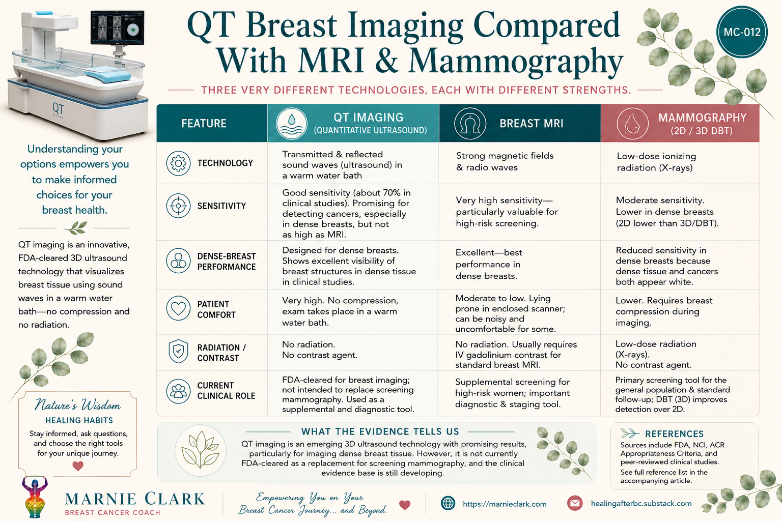
Always on the outlook for diagnostic tests that can help us understand what’s going on inside our breasts without the need for compression or ionizing radiation, today I am sharing with you Quantitative Transmission (QT) breast imaging.
Developed by QT Imaging Holdings, QT breast imaging is both radiation-free and compression-free. It uses sound wave technology to generate high-resolution, three-dimensional images of the breast, including maps showing how quickly sound travels through different tissues.
Imagine that – no breast compression and no ionizing radiation! But is it as good as mammography or MRI? And could it be particularly useful for women with dense breasts?
Let’s take a closer look.
How QT Imaging Works
QT imaging uses low-frequency ultrasound waves that travel through the breast. In transmission mode, the system measures the sound that passes through the breast and uses this information to create maps of speed, of sound, and other tissue characteristics. In reflection mode, the imaging provides additional information about tissue boundaries and structures.
The patient lies face down on a comfortable examination table, with the breast suspended in a warm water bath inside the scanner. The imaging arrays rotate around the breast to acquire the data, and the system then reconstructs the information into a three-dimensional image.
The scanning process is automated once the breast has been positioned, so there is no need for a technician as with conventional handheld ultrasound. It only takes between 10-15 minutes per breast to acquire the necessary imaging.
One important point: this doesn’t mean the entire appointment takes only 10-15 minutes – that’s just the image acquisition time for one breast – positioning and other parts of the examination add to the overall appointment time.
Is QT Imaging Better for Those With Dense Breasts?
This is one of the reasons QT imaging caught my attention.
Dense breast tissue contains a relatively high proportion of glandular and fibrous tissue and a lower proportion of fatty tissue. It is very common – in fact, the National Cancer Institute says that nearly half of women aged 40 and older have dense breasts.
Dense breast tissue matters for two reasons. First, having dense breasts is itself associated with a higher risk of breast cancer. Second, dense tissue can make cancers more difficult to see on a conventional mammogram.
Here’s a useful mental picture. On a mammogram, fatty tissue generally appears dark, while dense tissue appears white. Many breast cancers also appear white. So a cancer in a predominantly fatty breast may look a little like a white object against a dark background. But a cancer in a dense breast can be a little like looking for a white flower in a field of white flowers.
This doesn’t mean mammography is useless in dense breasts. Mammography (as much as we may not like it) remains an important and well-established screening tool, and 3D mammography (digital breast tomosynthesis, or DBT) can improve detection compared with conventional 2D mammography. But breast density can still make interpretation more difficult.
So Where Might QT Fit In?
QT imaging approaches the breast from a different direction. Rather than relying on x-rays to create the image, QT uses sound waves and measures characteristics such as speed of sound through different tissues, while also using reflected ultrasound information. This gives the system information about the internal structure and physical properties of the breast in three dimensions.
One early 2016 study compared QT images with breast anatomy and found that QT was able to identify major anatomical features, including glandular and ductal structures. The researchers also demonstrated that different breast tissues had distinguishable speed-of-sound characteristics.
This is particularly interesting in dense breasts because it suggests that QT may provide useful structural information within areas of dense tissue that might otherwise be difficult to distinguish in mammography.
However – and this is important – visualizing breast structures more clearly is not the same thing as proving that a technology detects more breast cancers.
QT Can Also Quantify Breast Density
Another interesting feature of QT is that it isn’t simply producing pictures. Because QT measures physical characteristics of tissue, researchers have investigated whether it can also provide a quantitative measurement of breast density.
A 2019 study found that QT-derived measurements of breast density were comparable with density measurements obtained from mammography with tomosynthesis. This is potentially useful because mammographic density is currently based on the appearance of tissue on an x-ray image, whereas QT provides an improved three-dimensional assessment.
This is still an area of research, but it adds another interesting dimension to what QT may eventually be able to tell us about breast tissue.
How Does QT Imaging Compare to Other Screening Methods?
Check out the infographic above to see how QT imaging differs from mammography and breast MRIs.
The comparison between QT imaging and mammography becomes particularly interesting when we look at an actual study involving breast radiologists.
A 2024 study published in Academic Radiology compared QT imaging with digital breast tomosynthesis (DBT), also known as 3D mammography.
Twenty-four breast radiologists interpreted 177 cases, testing whether QT was non inferior to DBT in overall diagnostic performance. The result was interesting: QT was found to be statistically non-inferior to DBT based on the study’s predefined criterion.
QT showed higher specificity than DBT (60.1% vs 37.2%), while DBT demonstrated higher sensitivity (85.2% vs 70.6%). These findings suggest that QT imaging has promising potential, particularly because of its ability to produce fewer false-positive recalls.
What Do Sensitivity and Specificity Actually Mean?
Let’s pause for a moment, because these two words can make a research paper sound much more complicated than it really is.
Sensitivity asks the question “How good is the test at finding the problem?” In other words, of all the people who really do have one of the target abnormalities (such as a tumor) how many does the test correctly identify?
In the study above:
QT sensitivity: 70.6%
DBT sensitivity: 85.2%
So within this particular study population, QT identified about 71 out of every 100 cases with the target abnormalities, whereas DBT identified about 85 out of every 100. DBT was therefore more sensitive in this study.
Specificity asks the question “How good is the test at recognizing what’s NOT a problem?” In other words, of all the cases that don’t have one of the target abnormalities, how many does the test correctly identify as not having an abnormality?
In this study:
QT specificity: 60.1%
DBT specificity: 37.2%
So QT was considerably more specific than DBT in this study. That means it was less likely to incorrectly flag a case as suspicious when the target abnormality wasn’t actually present.
Put very simply, sensitivity = finding the problem; specificity = correctly identifying what isn’t the problem.
In this particular study, DBT found more abnormalities, while QT generated fewer false-positive findings. That distinction is important. We shouldn’t turn the study into the simplistic claim that “QT is better than mammography” (a claim which I did find in an online article). The results suggest that the two technologies have different strengths.
What About Dense Breasts In This Study?
Now we come back to the question that initially caught my attention.
The researchers also looked specifically at women with dense breasts. QT performed well in this group, and the results were encouraging. However, the study was not large enough to prove that QT is better than 3D mammography at detecting breast cancer in women with dense breasts.
One particularly interesting finding was that QT’s ability to correctly identify non-suspicious breasts appeared to remain fairly consistent regardless of breast density. With 3D mammography, however, performance changed as breast density increased. This suggests that QT may have an advantage in maintaining consistent results in women with dense breasts, but this needs to be confirmed in larger studies.
That distinction is worth making because I don’t want you to walk away from this article thinking that women with dense breasts should abandon mammography in favor of QT. The science simply isn’t there yet.
QT Versus MRI
QT also has some practical advantages over breast MRI. A standard contrast-enhanced breast MRI generally requires an intravenous gadolinium-based contrast agent, and some people have reactions to this contrast agent. MRI also involves lying inside the scanner for a longer examination, which some people find uncomfortable or claustrophobic.
QT requires:
No ionizing radiation
No breast compression
No intravenous contrast injection
No enclosed MRI scanner
Instead, the breast is suspended in a warm water bath while the scanner acquires the images. That makes QT an intriguing option from a patient-comfort perspective. But MRI remains a very important tool because contrast-enhanced breast MRI has very high sensitivity, particularly in women at increased risk of breast cancer.
So again, we’re looking at different strengths rather than a simple winner and loser.
Who Might Be Interested in QT Imaging?
QT imaging is still an emerging technology, so I wouldn’t suggest that everyone should rush out and replace their usual breast screening with it. However, there are some women who may find this technology particularly interesting.
Women with dense breasts may be interested because QT uses a different imaging approach from mammography and can provide three-dimensional information about breast structures. Early research suggests that QT may be able to maintain consistent performance across different levels of breast density, although we still need larger studies to determine whether it actually detects more cancers in dense breasts.
Women who find mammography uncomfortable may be interested because QT does not compress the breast between plates. Instead, the breast is gently suspended in a warm water bath during the scan.
Women who prefer to avoid ionizing radiation, where clinically appropriate, may also be interested. QT uses sound waves rather than X-rays, so there is no ionizing radiation involved.
Women who cannot or do not want to undergo breast MRI may also want to learn more about QT. Unlike a standard contrast-enhanced breast MRI, QT does not require an intravenous contrast agent or lying inside an MRI scanner. However, this does not mean QT is equivalent to MRI, particularly for women who require the very high sensitivity that breast MRI can provide.
And finally, anyone interested in emerging breast-imaging technologies may want to keep an eye on QT. The technology is still being studied, but its combination of three-dimensional imaging, quantitative tissue measurements, no radiation and no compression makes it a fascinating development in breast imaging.
Two Limitations You Should Know About
There are a couple of important differences between QT and conventional breast imaging.
1. If a suspicious area is found, QT currently does not provide image-guided needle biopsy. The company QT Imaging says that a biopsy apparatus is being developed. That means QT is currently an imaging technology – if a suspicious finding requires tissue diagnosis, another method would still be needed to obtain the biopsy.
2. One reader raised an interesting question after reading this article: What about the tail of Spence and the lymph nodes under the arm? The tail of Spence is an extension of breast tissue that reaches upward and outward toward the armpit. Because this area lies close to the chest wall, imaging it completely can be challenging with some breast-imaging technologies.
Interestingly, QT Imaging has recently addressed this particular challenge. In March 2026, the company received FDA 510(k) clearance for an updated configuration of its Breast Acoustic CT scanner designed to improve visualization and expand coverage of posterior breast tissue near the chest wall. The company says the new configuration uses a tilted transmitter geometry to provide more complete visualization of this posterior breast anatomy.
However, I couldn’t find evidence showing that QT specifically provides complete visualization of the entire tail of Spence or that it is intended to replace other types of imaging of the axillary lymph nodes. If the lymph nodes need to be assessed, a healthcare professional may recommend targeted axillary ultrasound or another appropriate imaging examination.
So, for now, I would regard QT’s improved posterior-breast coverage as an encouraging development rather than saying that it completely solves the question of imaging the tail of Spence or axillary lymph nodes.
Regulatory Status for QT Imaging
In the United States, QT Imaging’s Breast Acoustic CT scanner has received multiple FDA 510(k) clearances.
The current QT Scanner 2000 Model A received another 510(k) clearance in March 2026. Importantly, the FDA’s current indication specifically states that the device is intended for breast imaging and is not intended to be used as a replacement for screening mammography
Internationally, QT Imaging has also announced regulatory clearances in the United Arab Emirates and Saudi Arabia.
QT Imaging is not currently listed in the Australian Register of Therapeutic Goods (ARTG), so it is not presently an established TGA-registered breast-imaging option in Australia.
The Bottom Line
I am excited that we are beginning to see breast imaging move beyond simply taking pictures of the breast. Technologies such as QT are exploring whether we can gather quantitative information about the tissue itself – how sound travels through it, how different structures are arranged, and potentially how those characteristics might change over time.
If you have dense breasts, I wouldn’t read this article and conclude that you should stop having mammograms and seek out QT imaging instead. Rather, I would encourage you to know your breast density, understand what your chosen imaging test can and cannot see, and have an informed conversation with your healthcare professional about whether additional imaging is appropriate for your individual risk profile.
You may find these articles helpful:
How to Detox After MRI Scans
Cone Beam Breast Computed Tomography – CBBCT – A More Comfortable and Accurate Alternative to Mammograms
References:
2016: Anatomy-Correlated Breast Imaging and Visual Grading Analysis Using Quantitative Transmission Ultrasound™ – https://pmc.ncbi.nlm.nih.gov/articles/PMC5056280/
2018: Quantitative transmission ultrasound tomography: Imaging and performance characteristics – https://pmc.ncbi.nlm.nih.gov/articles/PMC6041196/
2019: Quantitative Assessment of Breast Density: Transmission Ultrasound is Comparable to Mammography with Tomosynthesis – https://pubmed.ncbi.nlm.nih.gov/31645343/
2022: Comparison of the diagnostic performance of Magnetic Resonance Imaging (MRI), ultrasound and mammography for detection of breast cancer based on tumor type, breast density and patient’s history: A review – https://pubmed.ncbi.nlm.nih.gov/35148941/
2024: A Multireader Multicase (MRMC) Receiver Operating Characteristic (ROC) Study Evaluating Noninferiority of Quantitative Transmission (QT) Ultrasound to Digital Breast Tomosynthesis (DBT) on Detection and Recall of Breast Lesions – https://www.sciencedirect.com/science/article/pii/S107663322300716X
2026: QT Imaging Receives FDA Clearance for Its Breast Acoustic CT Scanner Enhanced Configuration to Improve Posterior Breast Imaging Coverage – https://ir.qtimaging.com/news-events/press-releases/detail/139/qt-imaging-receives-fda-clearance-for-its-breast-acoustic-ct-scanner-enhanced-configuration-to-improve-posterior-breast-imaging-coverage
National Cancer Institute article on Dense Breasts: https://www.cancer.gov/types/breast/screening/dense-breasts
USFDA article Understanding Breast Density: https://www.fda.gov/consumers/womens-health-topics/understanding-breast-density
About Marnie Clark

Hi I’m Marnie Clark, breast cancer survivor turned coach. I have 20 years of experience in natural medicine. In 2004/05 I battled breast cancer myself. You can see more about my journey on my page Breast Cancer Diary.
I’ve been healthy and recurrence-free since 2004 and in 2012 I became a Breast Cancer Coach because I became aware of the fact that whilst there is now a wealth of information on the Internet, much of it is confusing, conflicting, and sometimes just wrong!
So it is my duty to help you unconfuse and untangle all that information, and find what works for YOU.
GET MY BEST TIPS on healthy ways to beat breast cancer and prevent recurrences by signing up for my free e-newsletters and eBooks.
You can also “like” me on Facebook (Marnie Clark, Breast Health Coach) to get my inspirational snippets, news and updates. I promise to do my utmost to keep you informed and empowered on your healing journey… and beyond.
Subscribe On YouTube


Subscribe For Extra Support
Welcome to the Marnie Clark Breast Care Community. When you join my virtual family you will receive my informative newsletter PLUS my gift to you, these 2 eBooks valued at $47 to empower and support you on your breast cancer journey.
My subscribers also get a treasure trove of info on nutrition, supplements and lifestyle tips on surviving breast cancer.
“Thank you for the email and the meaningful words. I also wanted to thank you for being with me during my treatments. Your emails and posts not only gave me information but lots of courage.” Susan Z, Canada
0 Comments