 The Benefits of Thermography for Detecting Breast Cancer
Thermography, also known as thermal imaging, is an alternative method of screening for breast cancer that is completely safe, non-invasive, does not subject the breast to harmful radiation and doesn’t hurt at all!
Yet it is alarming how many people have never heard of it.
Mammography Is Not Saving Lives
If you read the research about breast cancer survival rates, everyone agrees that if breast cancer is detected in its earliest stages, 95 percent cure rates are possible.
One of the problems associated with mammography, ultrasound, MRI, and other commonly used imaging tools is that they rely primarily on finding a physical tumor. They cannot detect pre-cancerous changes in the breast.
Another problem with mammography is the repeated doses of radiation to the breast, which is a highly radio-sensitive organ. Each dose of radiation increases one’s risk for breast cancer. I read in two different places that a mammogram can expose you to approximately one thousand times the amount of radiation one might receive in a chest x-ray!
Further, if a tumor is present, the mechanical pressing upon it by traditional mammography methods can, some experts feel, rupture a cancerous tumor and seed the cancer cells throughout the breast where they can grow and spread.
A research paper published in September 2015 titled “Mammography Screening is Harmful and Should Be Abandoned” [1] makes it clear that despite decades of mammography screening, breast cancer mortality has not been reduced, rather it has led to millions of women being over-treated for supposedly early stage or stage zero breast cancer. In most cases, these women were offered toxic treatments they didn’t need, the result of which (in the case of chemotherapy and radiation) actually creates cancer stem cells, can alter benign cells to malignant cells, or transform cancer cells into much deadlier types.
Thermography Is Different
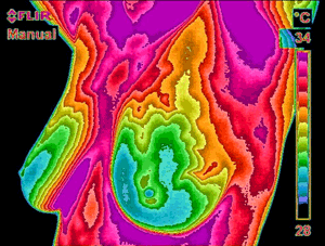
Thermography works quite differently, it investigates vascular changes in the breast, detects patterns of blood flow, inflammation and symmetrical changes.
You see, temperature changes in the breast can be an early indicator of the possible development of a cancerous tumor. Metabolic activity and vascular circulation in both pre-cancerous tissue and the area surrounding a developing breast tumor is almost always higher than in normal breast tissue. In an ever-increasing need for nutrients, tumors increase circulation in the area of the tumor by holding open existing blood vessels, opening dormant vessels, and creating new ones (called neoangiogenesis). This process causes an increase in the surface temperature of the breast and thermography can detect this.
Thermography uses ultra-sensitive medical infrared cameras and computer programs to detect, analyze, and produce high-resolution images of these temperature variations. By detecting tiny variations in blood vessel activity, thermal imaging may pick up a pre-cancerous state of the breast or the presence of an early tumor that is not yet large enough to be detected by physical examination, mammography, or other types of imaging.
Women who are on hormone replacement therapy, are nursing or who have fibrocystic, large, dense, or enhanced breasts can have problems with mammograms as these conditions make it quite difficult to see what is going on in the breast. With thermal imaging, however, these types of breast differences do not pose a problem since the technology utilized is quite different.
For those doctors who say that thermography isn’t proven and nothing more than quackery, research released in 2015 [2] indicated that “thermography may be useful in the initial screening and supplementation of diagnostic procedures due to its safety (its non-radiation properties), low cost and the good recognition of breast tissue disease.”
An older 2008 study [3] found that breast thermography had a 97 percent sensitivity rate in discovering malignancies, and that a digital infrared camera identified 58 out of 60 malignancies in breast tissue. The researchers involved in the study stated that DITI (Digital Infrared Thermal Imaging) was a valuable addition to mammography and ultrasound, especially in women with dense breast tissue.
The 7 Best Reasons for Using Thermography
- It doesn’t hurt!
- It is non-invasive, no squishing or damaging of breast tissue;
- The breast is not subjected to ionizing radiation every single year;
- Thermal imaging doesn’t cause cancer whereas mammography can;
- It takes 15 minutes;
- It is safe for pregnant women;
- Thermal imaging has the ability to detect a tumor developing when it is only the size of a pin head, some 3-5 years before a mammogram can see it.
What To Expect When You Get Thermography
This process may vary a little from place to place, but generally speaking, this is the routine to expect.
- You are invited to sit in a temperature-controlled room to allow your body to cool down. You will probably be asked to complete some health paperwork, including a health survey.
- For a breast thermogram, you will disrobe to the waist and be positioned in front of a thermal imaging camera. The technician will take digital pictures. You may be asked to plunge your hand into cold water on the side being photographed. The process takes between 5-15 minutes.
- Depending on the size of the facility and the staff, your pictures may either be read on the premises or sent out to a certified physician for analysis.
- You will receive a report of findings shortly thereafter and this can be used to help you and your medical practitioner to detemine any next steps.
Unfortunately, conventional medicine as a whole still has not accepted thermal imaging and few insurance companies will cover the cost of it, at least in the USA. The fee for first-time patients is around $200-300 in both the USA and Australia. Be that as it may, I believe it’s a much better method of discovering problems within the breast early on. It is well worth the price.
Please do not subject yourself to mammography screening as a “preventive measure”. By doing so, you are actually putting yourself right square into harm’s way. To find a certified thermography technician in your area, go to the American College of Clinical Thermology Inc (ACCT) website at www.thermologyonline.org. If you are located outside of the USA, just Google “breast thermography” and the name of your city/town/province to see if someone in your area performs this type of breast imaging.
References:
- Mammography screening is harmful and should be abandoned – http://jrs.sagepub.com/content/108/9/341.long
- Assessing the Potential of Thermal Imaging in Recognition of Breast Cancer – http://www.ncbi.nlm.nih.gov/pubmed/26745126
- Effectiveness of a Noninvasive Digital Infrared Thermal Imaging System in the Detection of Breast Cancer – http://www.ncbi.nlm.nih.gov/pubmed/18809055
- Does radiation-induced c-MYC amplification initiate breast oncogenesis? http://www.ncbi.nlm.nih.gov/pmc/articles/PMC4845163/
GET MY BEST TIPS on getting through breast cancer and preventing recurrences by signing up for my free e-newsletters and e-books on the right. You can also “like” me on Facebook (Marnie Clark, Breast Health Coach) to get my inspirational snippets, news and updates. I promise to do my utmost to keep you informed and empowered on your healing journey… and beyond.


I never knew that you could use thermal imaging to screen for breast cancer! I have an aunt that has breast cancer, so my family is more aware of getting our annual screenings. It’s also interesting that you point out that thermography doesn’t subject your body to harmful radiation, which I would imagine would be helpful if you were pregnant.
THANKS FOR THIS INFO. ITS A SHAME MORE WOMEN DON’T KNOW ABOUT IT.